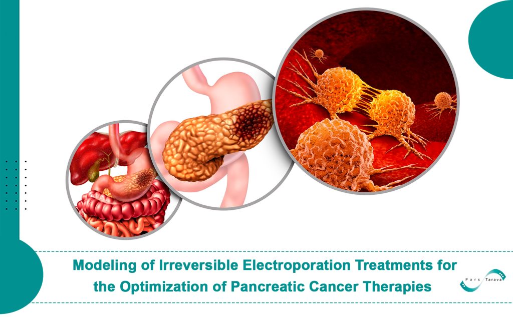
This article is a restructured and accessible adaptation of a scientific paper originally published in IFMBE Proceedings (Vol. 45) under the title “Modeling of Irreversible Electroporation Treatments for the Optimization of Pancreatic Cancer Therapies,” authored by E.L. Latouche and colleagues. The purpose of this rewrite is to present the core ideas and findings of the original study in a clear and reader-friendly format suitable for both medical professionals and a broader Persian-speaking audience interested in emerging cancer treatment technologies.
We’ve aimed to break down the essential components of the original research—such as an introduction to irreversible electroporation (IRE), its simulation process, and the evaluation of electric field and thermal effects in biological tissue. We’ve also included practical and clinical insights to bridge the gap between laboratory data and real-world application.
PARS Tarava, as the first Iranian company developing electroporation devices, is committed to making such advanced therapies accessible. By translating and explaining cutting-edge studies and presenting clinical examples, we aim to raise awareness about the potential of IRE in treating cancer and solid tumors. Our mission is to help medical professionals and patients benefit from this promising, non-invasive technology.
Understanding Pancreatic Cancer and Common Treatments
Pancreatic cancer is among the most aggressive and lethal types of cancer. The most prevalent form, pancreatic ductal adenocarcinoma (PDAC), originates in the cells lining the pancreatic ducts. Due to its late diagnosis in most cases, the 5-year survival rate remains under 5%.
Conventional treatment strategies include:
- Surgery: Feasible only in early stages; at best, it increases the 5-year survival rate to 20–25%.
- Chemotherapy and Radiation: Typically used to shrink tumors or improve quality of life; limited in long-term effectiveness.
- Combination Therapy: Chemotherapy with surgery or radiation can help in some cases, but remains limited in treating non-resectable tumors.
Emerging non-invasive methods, such as irreversible electroporation (IRE), offer new hope for treating advanced, non-operable tumors.
Introduction to Irreversible Electroporation (IRE)
Irreversible electroporation is a minimally invasive, non-thermal tumor ablation technique that uses high-voltage electrical pulses to create nanoscale pores in cell membranes, leading to cell death. This process delivers minimal energy to the tissue, avoiding substantial thermal damage. Unlike heat-based ablation methods like radiofrequency or laser, IRE preserves surrounding critical structures such as blood vessels and nerves.
This makes IRE particularly promising for treating tumors in complex and sensitive regions like the pancreas, where surgical options may be limited or too risky.
Why Simulation and Modeling Matter in IRE
Effective treatment planning in IRE requires an accurate understanding of how the electric field behaves in heterogeneous and anatomically complex tissue environments. The pancreas is especially challenging due to its proximity to vital structures and its irregular shape.
In this study, the authors created a 3D model of a patient’s pancreas, including the tumor and surrounding tissues, based on medical imaging (CT and MRI). This allowed them to predict treatment outcomes, such as ablation zones and heat distribution, with a high degree of precision.
Methodology
This section outlines the technical and step-by-step approach used to model and simulate irreversible electroporation. Key areas discussed include the 3D reconstruction from imaging data, software tools for meshing, the governing physical equations, and electrode configuration comparisons.
3D Reconstruction and Software Tools
Medical scans were obtained in axial, sagittal, and coronal planes, then processed using ParaView software to convert image data into a format suitable for finite element modeling. SolidWorks was used to design electrodes with a diameter of 19 AWG and 1.5 cm exposure length.
All geometries, including tumor, pancreas, and blood vessels, were meshed and imported into COMSOL Multiphysics for simulation.
Governing Equations
Two primary equations were used to simulate the behavior of the electric and thermal fields:
Laplace’s Equation
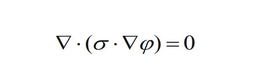
This equation governs the distribution of electric potential in the tissue. By solving it, the model predicts areas where the electric field exceeds the electroporation threshold (500 V/cm), identifying tissue likely to be ablated.
Pennes Bioheat Equation

This equation models thermal changes in biological tissue, accounting for blood perfusion, metabolic heat, and heat from electric pulses. It helps ensure that cell death results from electroporation, not thermal damage.
Electrode Configurations
Three arrangements were tested:
- Two electrodes spaced 2 cm apart
- Three electrodes spaced 1.25 cm apart
- Four electrodes in a 2×2 cm square configuration
Simulation Results
Tumor Coverage
The simulations showed that an electric field above 500 V/cm effectively covered much of the tumor area. The three-electrode configuration ablated approximately 73% of the tumor, outperforming the two-electrode setup (39%) and even the four-electrode one (59%).
This indicates that simply increasing the number of electrodes does not guarantee better results. Instead, optimal spacing and positioning of electrodes have a greater influence on treatment success.
Why Placement Matters More Than Quantity
Electric fields generated between electrodes follow specific spatial patterns. Poorly placed electrodes can lead to:
- Inadequate field coverage over parts of the tumor
- Unnecessary damage to healthy tissue
- Energy concentration in a small region, causing overheating
In contrast, the well-placed three-electrode arrangement created a more uniform and broader electric field, effectively ablating the tumor without involving much of the surrounding pancreas.
Thus, the design and spacing of electrodes based on tumor geometry are more critical than their count.
Thermal Effects
In the two-electrode model, thermal changes between electrodes were minimal, confirming that tissue damage primarily occurred via electroporation and not due to heat. The maximum recorded current across all models was 40.1 A.
Benefits of Pre-Treatment Simulation
The simulation model enables physicians to evaluate different electrode setups before the procedure. It helps avoid accidental contact with blood vessels and optimizes the ablation volume while minimizing harm to healthy tissue. This leads to safer and more effective treatment outcomes.
Conclusion and Future Directions
This study demonstrates how computational modeling based on patient-specific imaging can guide optimal IRE treatment for pancreatic cancer. Although some biological factors—such as changes in tissue conductivity or the effect of endothelial layers—were excluded in this model, future work aims to incorporate them.
More accurate simulations that reflect dynamic changes during electroporation could help fine-tune treatment protocols and improve patient outcomes. Measuring real-time conductivity and integrating both electrical and thermal models will offer deeper insights into the mechanism of cell death and treatment boundaries.
Irreversible electroporation is emerging as a powerful tool for treating non-resectable tumors. Advanced modeling, such as the one shown in this study, paves the way for its optimized and widespread use.